
3D In Vivo Optical Imaging System
3D In Vivo Optical Imaging System
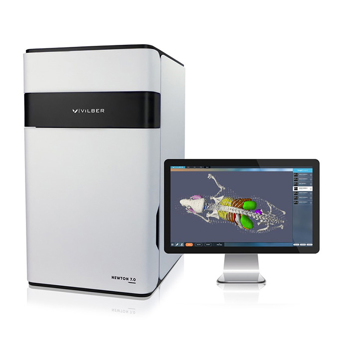
Model:Newton 7.0 FT-500
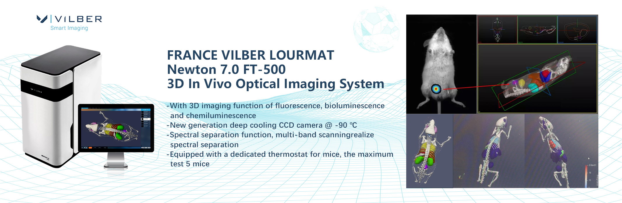
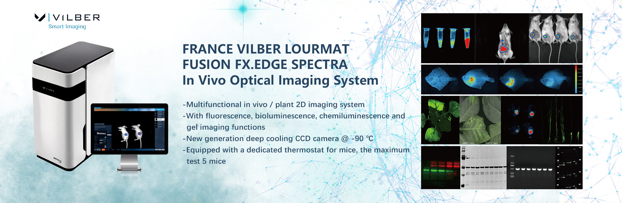
Newton 7.0FT-500The 3D small animal in vivo imaging system has the functions of fluorescence, bioluminescence, and chemiluminescence 2D/3D imaging.Thesystem combines the characteristics of high sensitivity, high optical signal acquisition capability, and user-friendly operation interface.
Using a new generation of deep cooling CCD camera, dark current 0.0001e/p/s@-90℃, cooling speed is faster and life is longer;
The f0.7 super lens greatly increases the amount of light entering per unit time, thereby effectively shortening the exposure time, and is especially suitable for bioluminescence imaging of Luc reporter gene detection
Fluorescence imaging uses double-sided high-intensity pulsed LED array scanning light source, multi-channel, and the excitation intensity is higher than that of halogen lamps. Based on scanning mode, excitation uniformity ≥99%;
10-bit emission filter wheel, equipped with 9 dedicated narrow-wave filters to meet the detection needs of various dyes;
Covers the spectral separation function, through multiple scanning of multiple different wavelength lasers, spectral splitting;
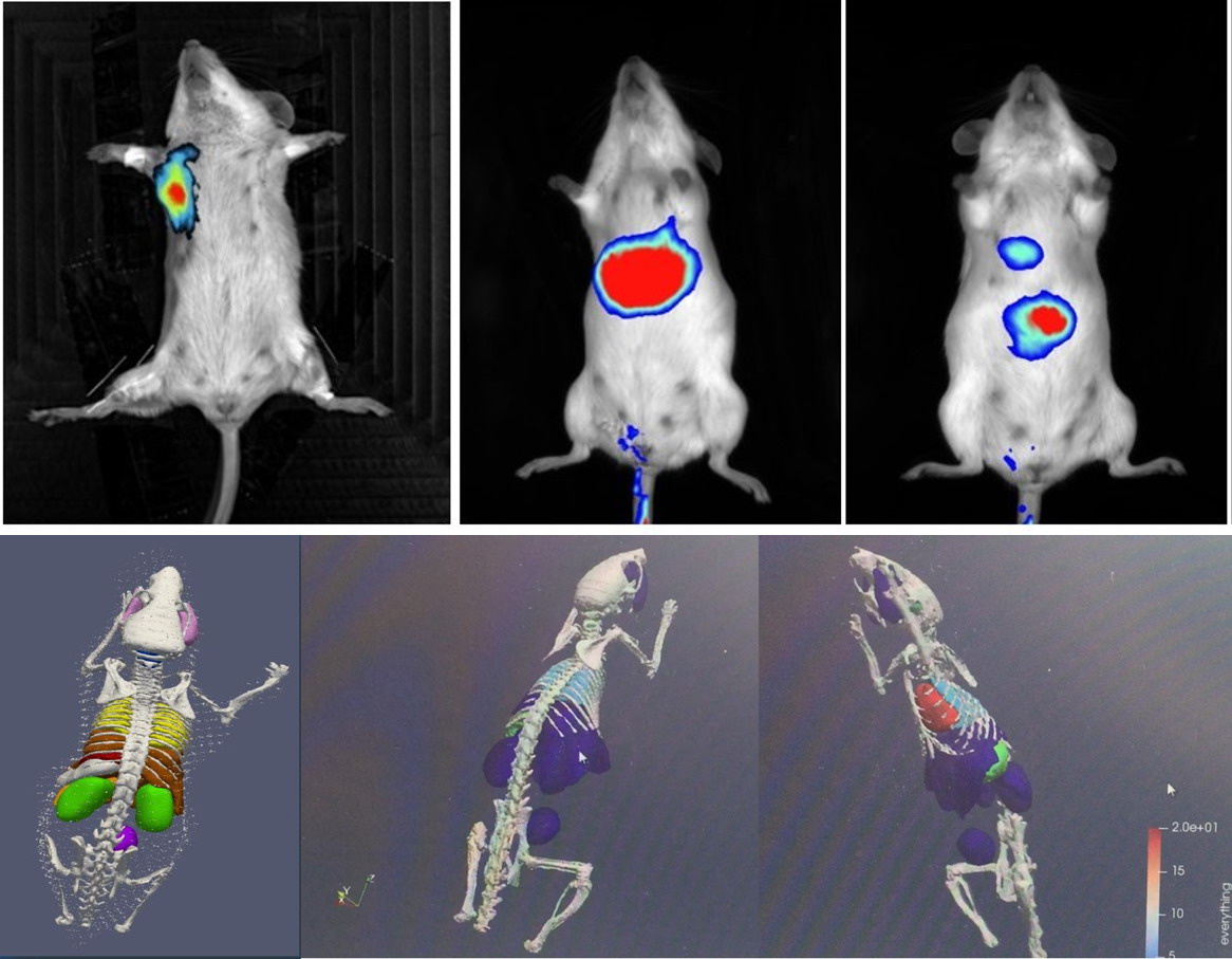
With topology and tomography technology, it realizes optical three-dimensional imaging and accurately locates the signal source;
It has a CCD camera that can be raised and lowered, and a stage that can move along the X and Y axes to achieve three-dimensional control, especially to meet the needs of local imaging of mice and obtain more details;
Equipped with special thermostat for mice, compatible with mouse anesthesia system, the maximum flux can reach 5 mice.
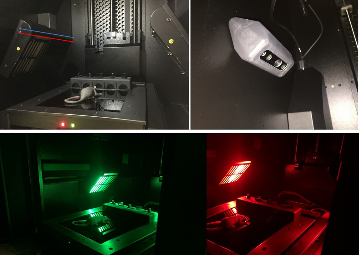
Figure 1.Newton 7.0 FT-500 double-sided array scanning light source
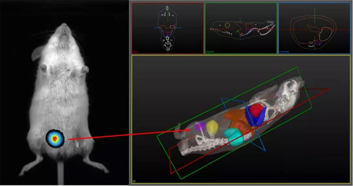
Figure 2. Newton 7.0 FT-500 topology and tomography technology to achieve optical 3D imaging and accurate positioning of the signal source
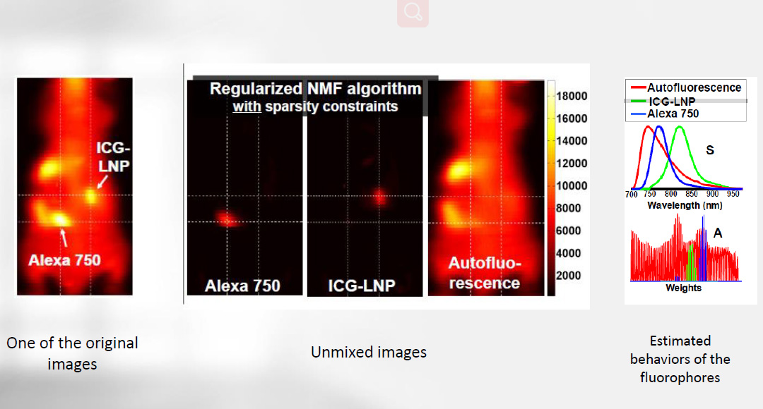
Figure 3. Newton 7.0 FT-500 spectral separation function, splitting the target fluorescence signal from autofluorescence
CONTACT
|
FEEDBACK
 *
*
Submit
|
SERVICE HOTLINE
+86-18516393082 
|


Copyright © Jinghe Imaging Science (Shanghai) Co., Ltd. All rights reserved. 沪ICP备20010438号
Do not embezzle our pictures and articles, otherwise we will investigate their legal responsibility.